|
|
| |
|
 |
|
| |
 HOME > Functional Assays > Cell Viability Assay |
|
|

|
Cell Viability Assay Kit *Blue |
| |
|
|
|
|
|
 |
코 드 |
: 22785 |
|
|
단 위 |
: 5 plates |
|
|
공 급 원 |
: AAT Bioquest |
|
|
가 격 |
: 문의 |
|
다운로드 |
|
|
|
|
|
|
|
| |
|
|
|
| |
 |
|
 |
| |
|
|
|
| |
 |
| |
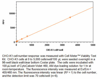
Cell Viability Assay Kit
Cell Meter™ Cell Viability Assay Kit *Blue Fluorescence*
This kit uses a proprietary dye that gets enhanced fluorescence upon entering into live cells. The dye is a hydrophobic compound that easily permeates intact live cells. The hydrolysis of the weakly fluorescent substrate by intracellular esterases generates a strongly fluorescent hydrophilic product that is well-retained in the cell cytoplasm. The esterase activity is proportional to the number of vial cells, and thus directly related to the fluorescence intensity of the product generated from the esterase-catalyzed hydrolysis of the fluorogenic substrate. Cells grown in black-walled plates can be stained and quantified in less than two hours.
The assay is more robust than the tetrazolium salt or Alarmar Blue™-based assays. It can be readily adapted for high-throughput assays in a wide variety of fluorescence platforms such as microplate assays, immunocytochemistry and flow cytometry. It is useful for a variety of studies, including cell adhesion, chemotaxis, multidrug resistance, cell viability, apoptosis and cytotoxicity. It is suitable for proliferating and non-proliferating cells, and can be used for both suspension and adherent cells. Using 100μl of reagents per well in a 96-well format, this kit provides sufficient reagents to perform 500 assays. Using 25μl of reagents per well in a 384-well format, this kit provides sufficient reagents to perform 2,000 assays.
Key Features
Increased Signal Intensity : Higher maximum signal with lower variation across the plate.
Rapid Dye Loading : Dye loading at RT for 30 min to 1 hr.
Convenient and Robust : Formulated to have minimal hands-on time.
Versatile applications : Compatible with many cell lines and targets
Kit Components
Component A : CytoCalcein™ Violet 460, AM 5 vials, lyophilized
Component B : DMSO 1 vial (200 μL)
Component C : Assay Buffer 1 bottle (50 mL)
Brief Summary
→ Prepare cells with test compounds
→ Add the same volume of dye-loading solution (100 μL for 96-well plate or 25 μL for 384-well plate)
→ Incubate at room temperature or 37℃ for 1 hr
→ Read Fluorescence at Ex/Em = 405/460 nm
References
1. Zibek S, Stett A, Koltay P, Hu M, Zengerle R, Nisch W, Stelzle M. (2006) Localized functional chemical stimulation of TE 671 cells cultured on nanoporous membrane by calcein and acetylcholine. Biophys J.
2. Klesius PH, Evans JJ, Shoemaker CA, Pasnik DJ. (2006) A vaccination and challenge model using calcein marked fish. Fish Shellfish Immunol, 20, 20.
3. Bratosin D, Mitrofan L, Palii C, Estaquier J, Montreuil J. (2005) Novel fluorescence assay using calcein-AM for the determination of human erythrocyte viability and aging. Cytometry A, 66, 78.
4. Schoonen WG, Westerink WM, de Roos JA, Debiton E. (2005) Cytotoxic effects of 100 reference compounds on Hep G2 and HeLa cells and of 60 compounds on ECC-1 and CHO cells. I mechanistic assays on ROS, glutathione depletion and calcein uptake. Toxicol In Vitro, 19, 505. |
| |
|
| |
 |
| |
22785
|
Product Code |
Unit |
Price |
Availability |
|
22785 |
5 plates |
|
|
|
| |
|
|