| |
ADP Assay kit
PhosphoWorks™ Fluorimetric ADP Assay Kit *Red Fluorescence*
ADP is involved in many biological reactions such as protein kinases. Our ADP assay kit can be used for assaying protein kinase reactions universally by monitoring ADP formation, which is directly proportional to enzyme phosphotransferase activity and is measured fluorimetrically. This kit provides a fast, simple and homogeneous assay for measuring ADP formation or depletion. The assay is continuous, and can be easily adapted to automation. The kit is convenient, requiring minimal hands-on time. Protein kinases are of interest to researchers involved in drug discovery due to their broad relevance to diseases such as cancer and other proliferative diseases, inflammatory diseases, metabolic disorders and neurological diseases. Most of commercial protein kinase assay kits are either based on monitoring of phosphopeptide formation or ATP deletion. Our ADP assay kit is more robust for assaying protein kinases than most of commercial kinase assay kits.
Key Features
Universal : Can be used for any kinases that used ATP as phosphate donor.
Continuous : Easily adapted to automation with no mixing or separation protocols.
Convenient : Formulated to have minimal hands-on time.
Non-Radioactive : No special requirements for waste treatment.
Use of Native substrates : Substrates can be proteins, peptides or sugars.
Large Range of ATP Tolerance : ATP can be used from1-300 μM.
Non-Antibody-Based : No antibody is used in the kit.
Kit Components
Component A : ADP Sensor Buffer 1 vial (2 ml)
Component B : ADP Sensor (Light-sensitive) 1 vial (1 ml)
Component C : ADP Standard 1 vial
Component D : ADP Assay Buffer 1 vial (5 ml)
Brief Summary
→ Run kinase reaction (20 μL)
→ Add component A (20 μL)
→ Add component B (10 μL)
→ Incubate at room temperature for 15 min-1 hr → Read fluorescence at Ex 540 nm/Em 590 nm
→ Read fluorescence at Ex 540 nm/Em 590 nm
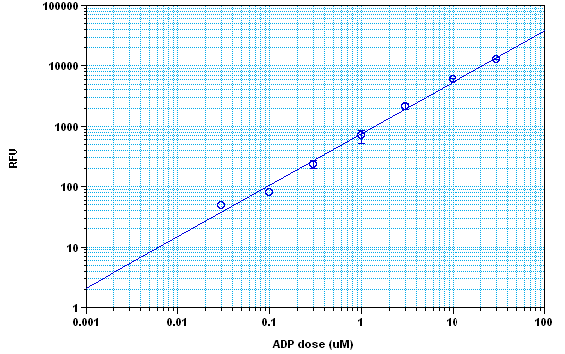
 Figure1. ADP dose response on 384-well black plate using a Gemini fluorescence microplate reader (Molecular Devices) measured with the PhosphoWorksTM Fluorimetric ADP Assay Kit. As low as 0.3 μM ADP can be detected with 15, 30 minutes and 1 hour incubation time (Z’ factor =0.65).
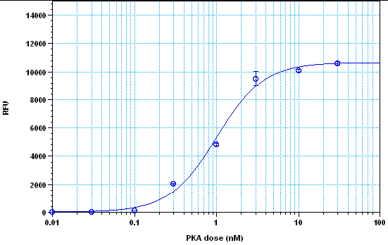
 Figure 2. Protein kinase A detection with the PhosphoWorksTM Fluorimetric ADP Assay Kit. The kinase was incubated in the presence of ATP and kemptide peptide substrate for 30 minutes, and ADP generation was detected at 30 min incubation time using the PhosphoWorksTM Fluorimetric ADP Assay Kit.
References
1. Cohen P (2000). The regulation of protein function by multisite phosphorylation—a 25 year update. Trends Biochem Sci 25, 596-601.
2. Whitmarsh AJ and Davis RJ (2000). Regulation of transcription factor function by phosphorylation. Cell Mol Life Sci 57, 1172-83.
3. Hunter T (1998). The role of tyrosine phosphorylation in cell growth and disease. Harvey Lect 94, 81-119.
4. McCubrey JA, et al. (2000). Serine/threonine phosphorylation in cytokine signal transduction. Leukemia 14, 9-21.
5. Salli U, et al. (2000). Phosphorylation of myristoylated alanine-rich C kinase substrate (MARCKS) protein is associated with bovine luteal oxytocin exocytosis. Biol Reprod 63, 12-20.
6. Parekh DB, et al. (2000). Multiple pathways control protein kinase C phosphorylation. Embo J 19, 496-503. |


